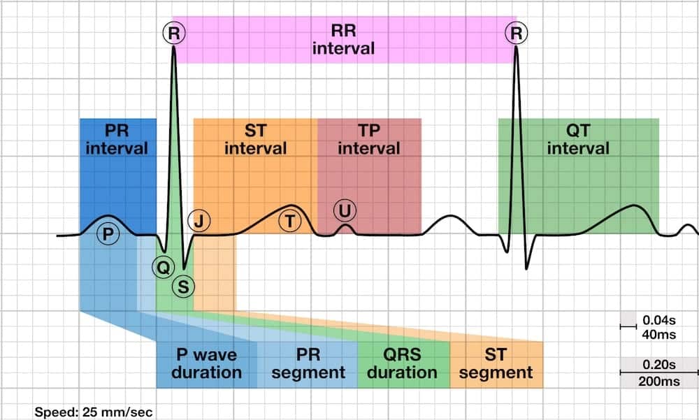
U wave Overview
The U wave is a small (0.5 mm) deflection immediately following the T wave
- U wave is usually in the same direction as the T wave.
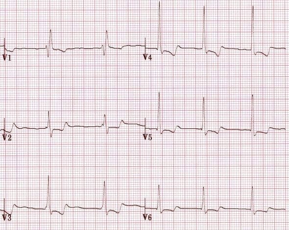
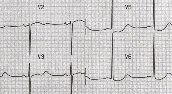
- U wave is best seen in leads V2 and V3.
Source of the U wave
The source of the U wave is unknown. Three common theories regarding its origin are:
- Delayed repolarisation of Purkinje fibres
- Prolonged repolarisation of mid-myocardial “M-cells”
- After-potentials resulting from mechanical forces in the ventricular wall
Features of Normal U waves
- The U wave normally goes in the same direction as the T wave
- U -wave size is inversely proportional to heart rate: the U wave grows bigger as the heart rate slows down
- U waves generally become visible when the heart rate falls below 65 bpm
- The voltage of the U wave is normally < 25% of the T-wave voltage: disproportionally large U waves are abnormal
- Maximum normal amplitude of the U wave is 1-2 mm
Abnormalities of the U wave
- Prominent U waves
- Inverted U waves
Prominent U waves
U waves are described as prominent if they are
- >1-2mm or 25% of the height of the T wave.
Causes of prominent U waves
Prominent U waves most commonly found with:
Prominent U waves may be present with:
- Hypocalcaemia
- Hypomagnesaemia
- Hypothermia
- Raised intracranial pressure
- Left ventricular hypertrophy
- Hypertrophic cardiomyopathy
Drugs associated with prominent U waves:
- Digoxin
- Phenothiazines (thioridazine)
- Class Ia antiarrhythmics (quinidine, procainamide)
- Class III antiarrhythmics (sotalol, amiodarone)
Note many of the conditions causing prominent U waves will also cause a long QT.
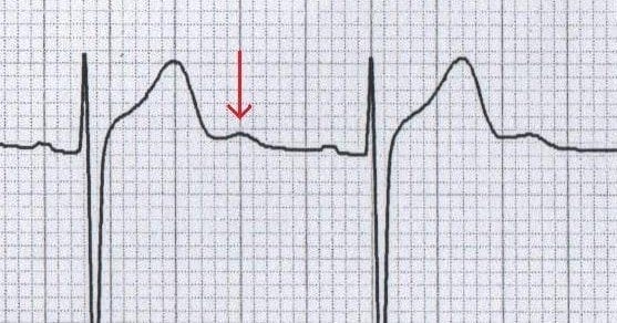
Prominent U waves due to sinus bradycardia
U waves associated with hypokalaemia

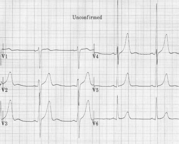
U waves associated with left ventricular hypertrophy
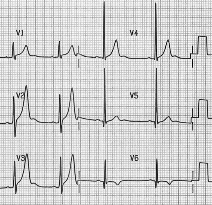
U waves associated with digoxin use
U waves associated with quinidine use
Inverted U waves
- U-wave inversion is abnormal (in leads with upright T waves)
- A negative U wave is highly specific for the presence of heart disease
Common causes of inverted U waves
- Coronary artery disease
- Hypertension
- Valvular heart disease
- Congenital heart disease
- Cardiomyopathy
- Hyperthyroidism
In patients presenting with chest pain, inverted U waves:
- Are a very specific sign of myocardial ischaemia
- May be the earliest marker of unstable angina and evolving myocardial infarction
- Have been shown to predict a ≥ 75% stenosis of the LAD / LMCA and the presence of left ventricular dysfunction
Unstable angina
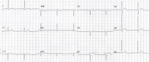
- Inverted U waves in a patient with unstable angina. Reproduced from Girish et al.
Inverted U waves in Prinzmetal angina

NSTEMI
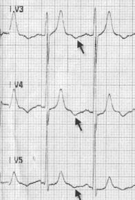
- Note the subtle U-wave inversion in the lateral leads (I, V5 and V6) in this patient with a NSTEMI; these were the only abnormal findings on his ECG.
No comments:
Post a Comment