NOVEMBER 1, 2013
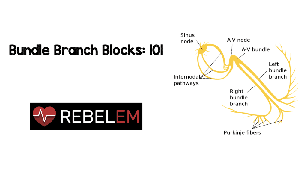
Bundle Branch Blocks: 101
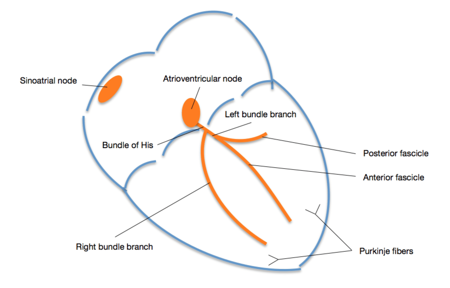
Written by Salim Rezaie REBEL EM Medical Category: Cardiovascular Recently, I have been asked by several students at my home institution (UTHSC at San Antonio) to help them understand bundle branch blocks. This is different than some of my usual posts because it is meant to be more educational than evidence based. So here we go. The normal conduction system of the healthy heart is shown to the right. If there is a delay or block in the left or right bundle, depolarization will take longer to occur. Therefore we get a widened QRS (>0.12 sec or >3 small boxes).
Conceptual Tip
One thing to remember to make this easier on yourself is that an “S Wave,” essentially means depolarization is going away from something and an “R Wave,” means depolarization is going toward something. As you continue to read this post just remember S = Away and R = Toward.
Is it LBBB or RBBB?
Once you have identified that your QRS is wide go to lead V1. If the “terminal force” of the QRS is above the baseline (big R wave) you have a RBBB. If the “terminal force” of the QRS is below the baseline (big S wave) you have a LBBB.
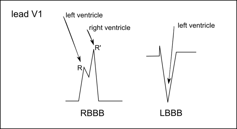
Image from ECGPedia.org
Why does a LBBB have a “big S Wave” and RBBB a “big R Wave” in lead V1?
- In RBBB, the last depolarization to occur is in the right ventricle therefore the left ventricle depolarizes first, which means the conduction is moving toward V1 (Left to Right).
- In LBBB, the last depolarization to occur is in the left ventricle therefore the right ventricle depolarizes first, which means the conduction is moving away from V1 (Right to Left).
- Remember the right side of the heart sits closer to the chest wall than the left side of the heart
Are there more specific criteria for RBBB and LBBB?
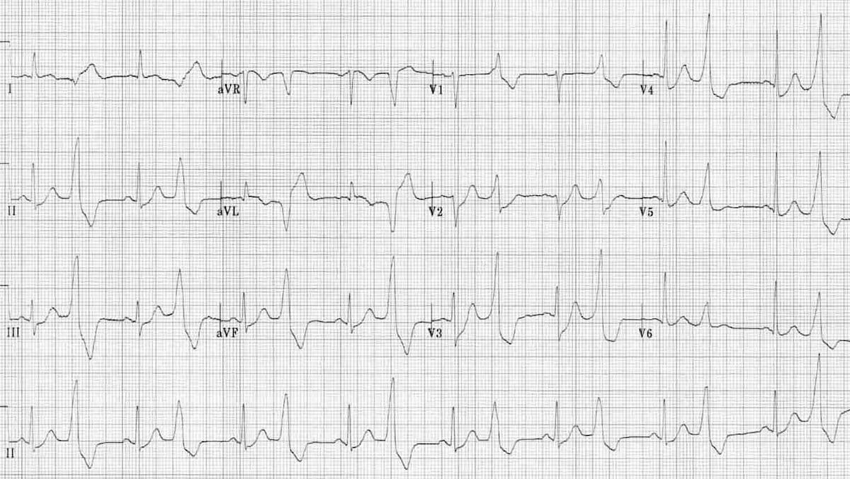
- RBBB
- QRS duration >0.12 seconds
- Slurred S wave in lead I, aVL, V5, and V6 (Depolarization moving away from these leads)
- RSR’ in V1 and V2 with R’ > R (Depolarization moving toward these leads)
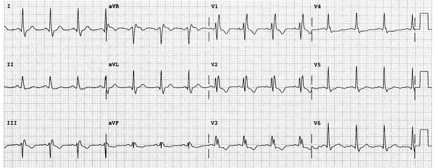
ECG from Dr. Smith’s ECG Blog
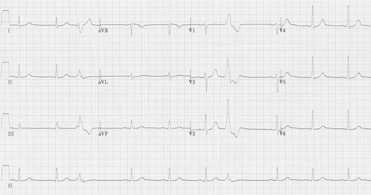
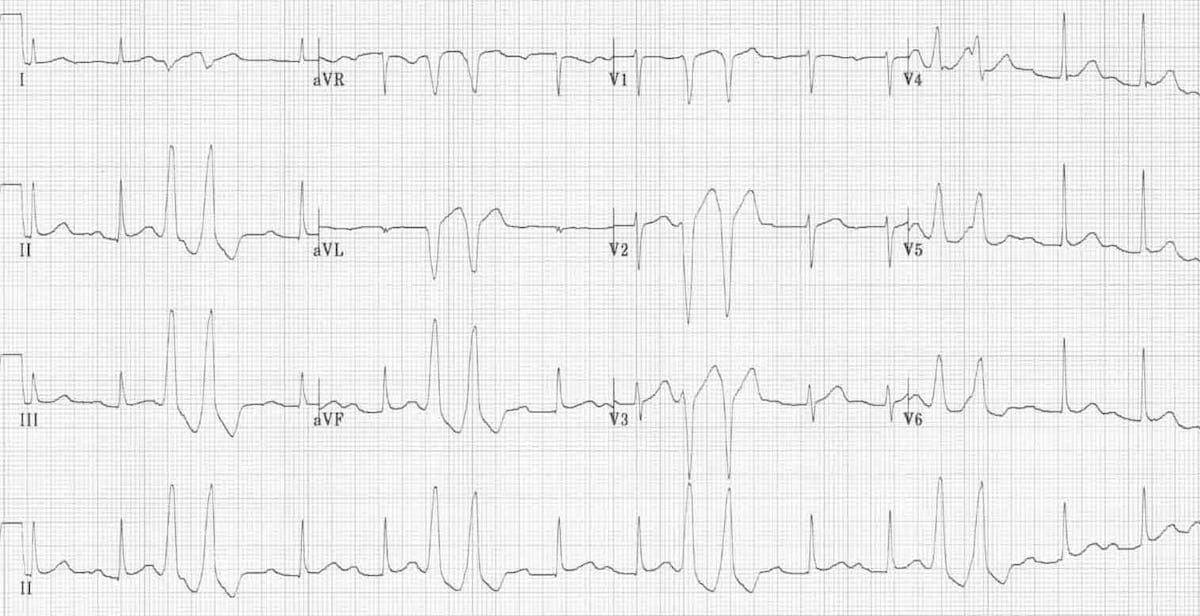
- LBBB
- QRS duration >0.12 seconds
- Broad monomorphic R waves in I, aVL, V5, and V6 (Depolarization moving toward these leads)
- Broad, dominant, monomorphic S wave in V1 and V2 (Depolarization moving away from these leads)
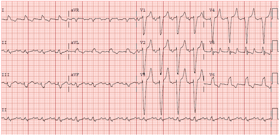
ECG from Dr. Smith’s EGG Blog
Left bundle with anterior and posterior fascicle
I wish I could say that it is just that easy, but it gets more complicated. The left bundle actually has an anterior and a posterior fascicle. This changes the morphology and the axis of the ECG. I will try and go through this to help simplify the ECG as we did with LBBB and RBBB.
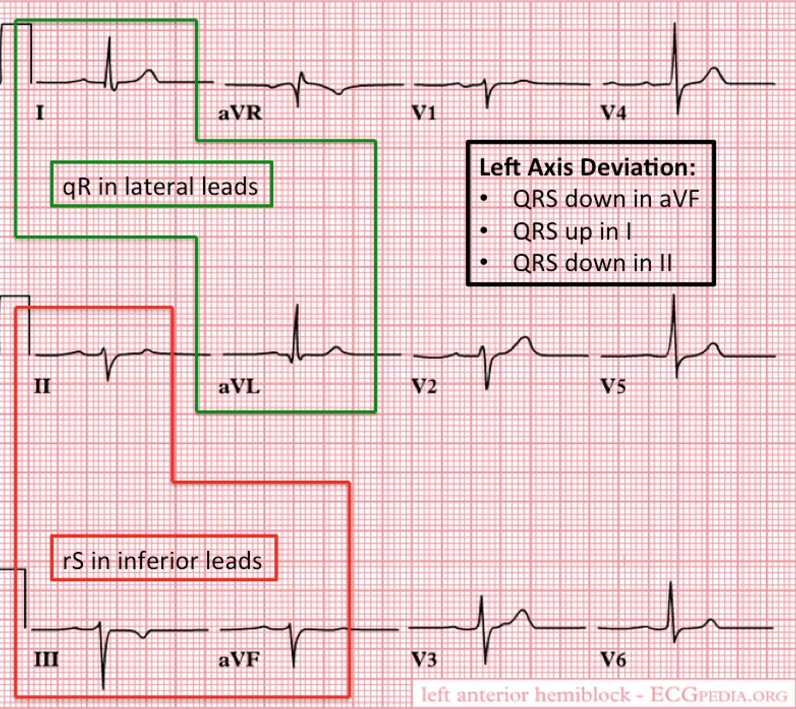
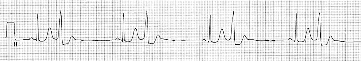
Left anterior fascicular block (LAFB)
Lets start with LAFB. If the left anterior fascicle is blocked, then depolarization in the left ventricle will go toward the lateral leads (I and aVL) and away from the inferior leads (II, III, and aVF). This means you would expect to see a bigger R wave in leads I and aVL and a bigger S wave in leads II, III, and aVF.
What are the criteria for LAFB?
- Slightly prolonged QRS duration (Not quite 120 msec or < 3 small boxes)
- Left axis deviation
- qR complex in leads I and aVL (Depolarization going towards these leads)
- rS complex in leads II, III, and aVF (Depolarization going away from these leads)
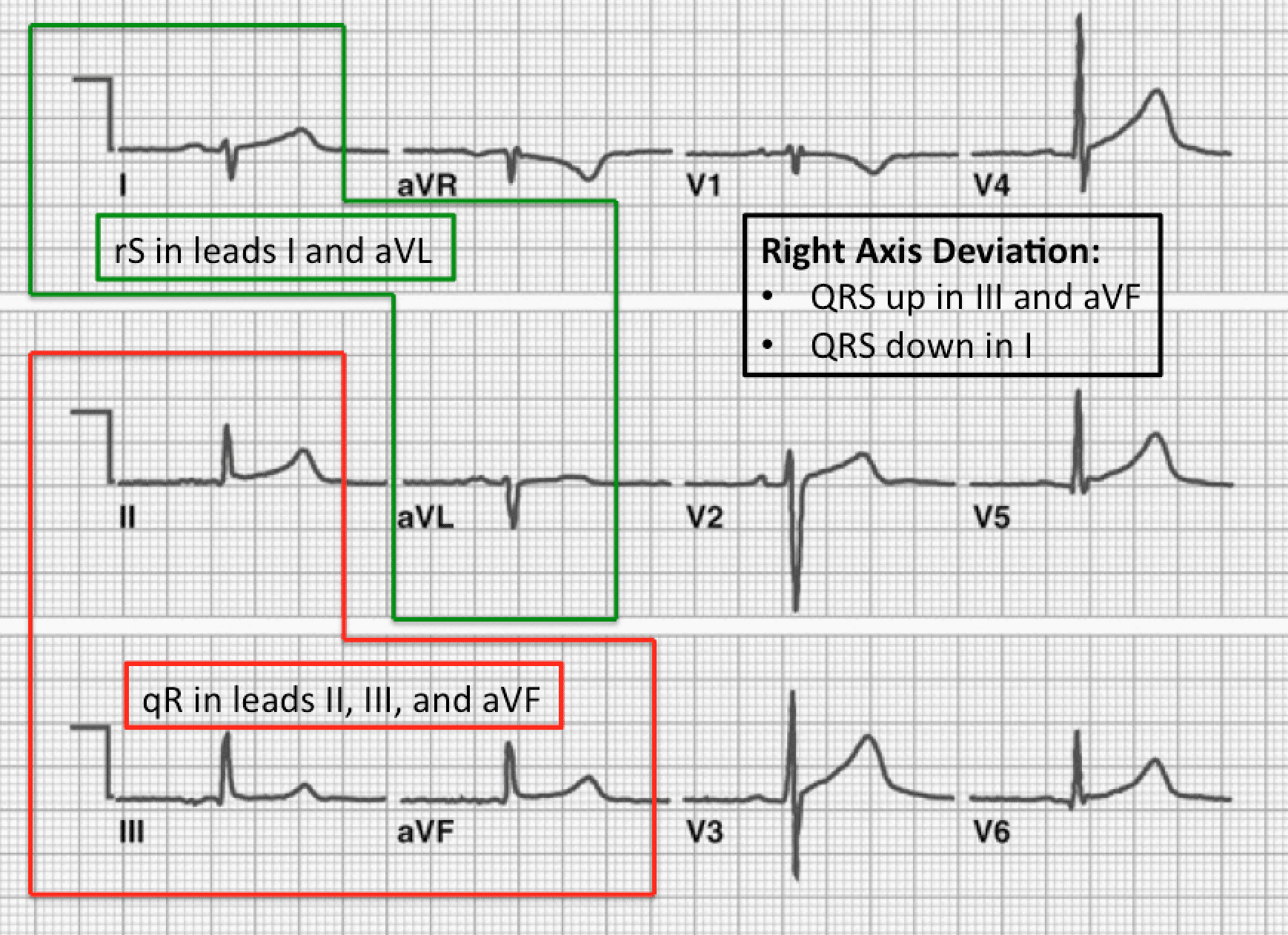
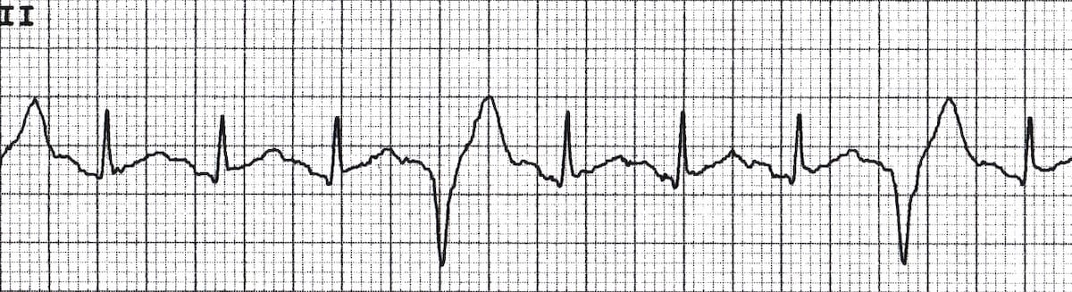
Left Posterior Fascicular Block (LPFB)
Lets now move on to LPFB. In general it is rare to find LPFB in isolation, because it is typically seen with RBBB. For the sake of explanation, if the posterior fascicle is blocked in isolation, then depolarization in the left ventricle will go toward the inferior leads (II, III, and aVF) and away from the lateral leads (I and aVL). This means you would expect to see a bigger R wave in leads II, III, and aVF and a bigger S wave in leads I and aVL.
What are the criteria for LPFB?
- Slightly prolonged QRS duration (Not quite 120 msec or < 3 small boxes)
- Right axis deviation
- qR complex in leads II, III, and aVF (Depolarization going towards these leads)
- rS complex in leads I and aVL (Depolarization going away from these leads)
- Absence of right ventricular hypertrophy or prior lateral myocardial infarction
Hopefully, this helps simplify bundle branch blocks and how to read them on ECGs. Just remember:
S wave = depolarization away from leads
R wave = depolarization towards leads
Cite this article as: Salim Rezaie, "Bundle Branch Blocks: 101", REBEL EM blog, November 1, 2013. Available at: https://rebelem.com/bundle-branch-blocks101/. The following two tabs change content below.Creator & Founder of REBEL EM


Sorry, the comment form is closed at this time.