Hypokalaemia
Hypokalaemia is defined as a serum potassium level of < 3.5 mmol/L. ECG changes generally do not manifest until there is a moderate degree of hypokalaemia (2.5-2.9 mmol/L). The earliest ECG manifestation of hypokalaemia is a decrease in T wave amplitude.
ECG features of hypokalaemia (K < 2.7 mmol/L)
- Increased P wave amplitude
- Prolongation of PR interval
- Widespread ST depression and T wave flattening/inversion
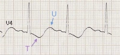
- Prominent U waves (best seen in the precordial leads V2-V3)
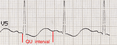
- Apparent long QT interval due to fusion of T and U waves (= long QU interval)
With worsening hypokalaemia…
- Frequent supraventricular and ventricular ectopics
- Supraventricular tachyarrhythmias: AF, atrial flutter, atrial tachycardia
- Potential to develop life-threatening ventricular arrhythmias, e.g. VT, VF and Torsades de Pointes
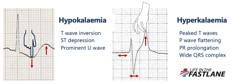
The push-pull effect
- Hypokalaemia creates the illusion that the T wave is “pushed down”, with resultant T-wave flattening/inversion, ST depression, and prominent U waves
- In hyperkalaemia, the T wave is “pulled upwards”, creating tall “tented” T waves, and stretching the remainder of the ECG to cause P wave flattening, PR prolongation, and QRS widening
Pathophysiology
Potassium is vital for regulating the normal electrical activity of the heart. Decreased extracellular potassium causes myocardial hyperexcitability with the potential to develop re-entrant arrhythmias.
| Degree of hypokalaemia | Potassium level (mmol/L) |
| Mild | 3.0 – 3.4 |
| Moderate | 2.5 – 2.9 |
| Severe | ≤ 2.4 |
Handy tips
- Hypokalaemia is often associated with hypomagnesaemia, which increases the risk of malignant ventricular arrhythmias
- Check both potassium and magnesium levels in any patient with an arrhythmia
- Replace potassium to ≥ 4.0 mmol/L and magnesium to ≥ 1.0 mmol/L to stabilise the myocardium and protect against arrhythmias – this is standard practice in most CCUs and ICUs
ECG Examples
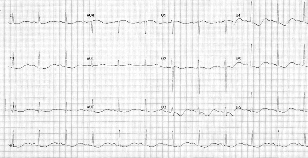
Example 1
Hypokalaemia:
- Widespread ST depression and T wave inversion
- Prominent U waves
- Long QU interval
This patient had a serum K+ of 1.7
Example 2
Hypokalaemia
- ST depression and T wave inversion best noted in inferior leads
- Prominent U waves
- Long QU interval
The serum K+ was 1.9 mmol/L.
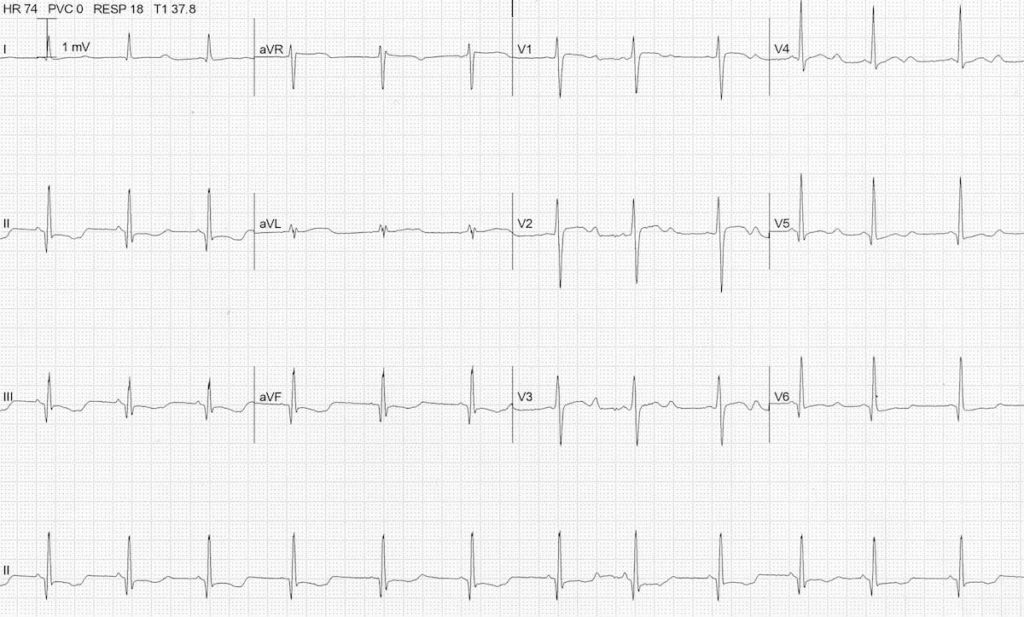
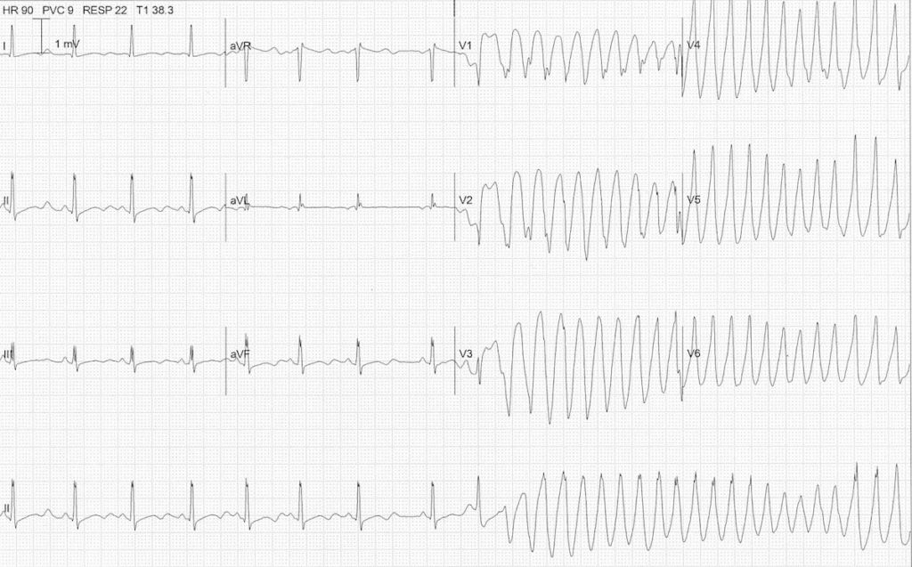
Example 3
Hypokalaemia causing Torsades de Pointes
- Another ECG from the same patient
- Note the atrial ectopic causing ‘R on T’ (or is it ‘R on U’?) that initiates the paroxysm of TdP
No comments:
Post a Comment